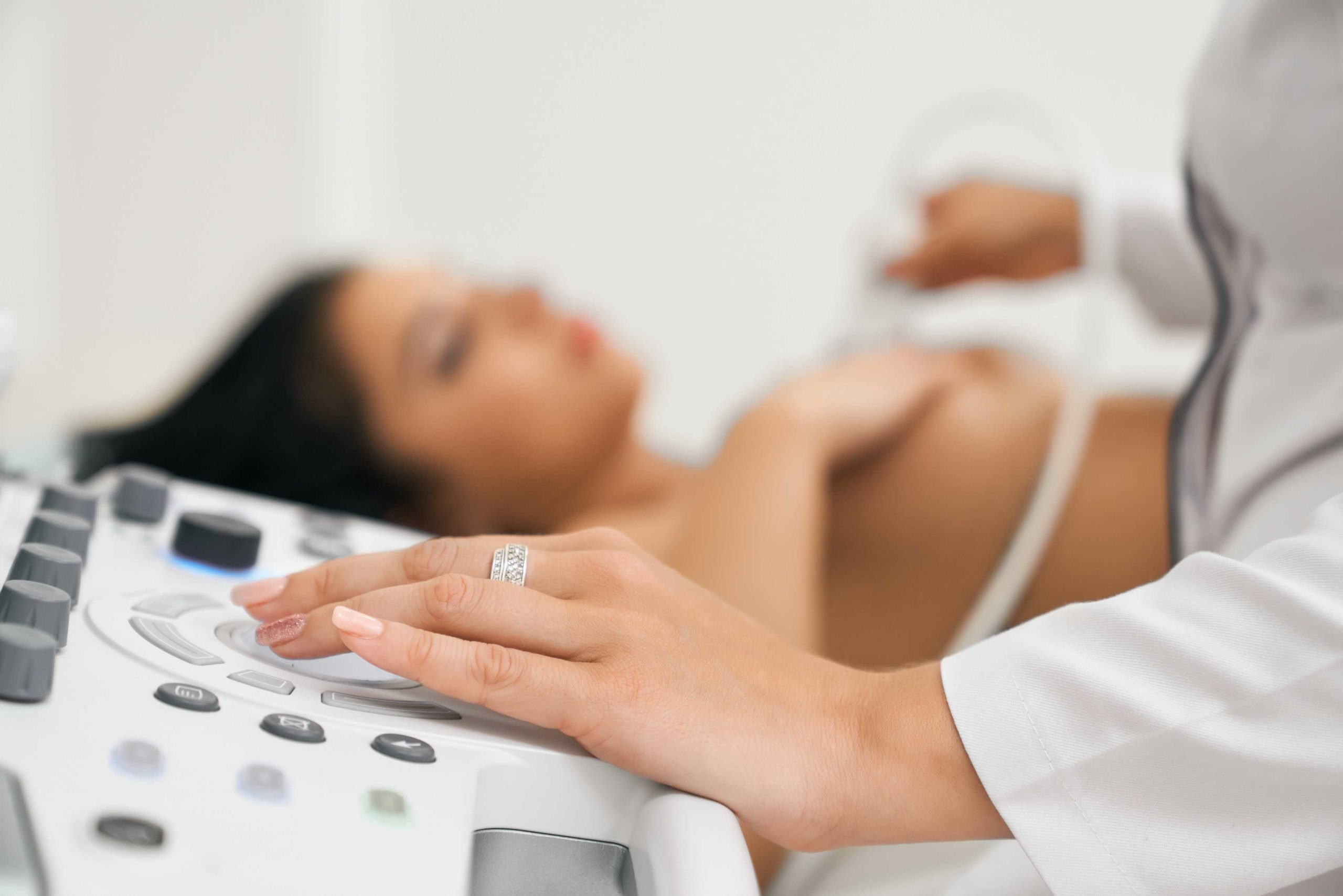
Breast ultrasounds are simple, noninvasive procedures that use sound waves and create images of the breast tissues. They are painless and safe from radiation because the sound wave technology bounces or echoes off the various breast tissues and forms visual images.
Breast ultrasounds can be used to detect:
In many cases, patients who need imaging choose breast ultrasounds because they are relatively quick and easy and use no radiation.
Ultrasound imaging is a common imaging technique used to monitor pregnancies, so the process here is very similar, but for a different part of the body. Patients lie down on their backs on a bed with their arms raised up over their heads. A member of our imaging staff applies a clear gel that helps them glide the transducer around the breast tissues. The transducer is connected to a computer screen where the sound signals are converted into real-time images.
Breast ultrasounds are performed by trained breast radiologists and completed within 30 minutes. The reports are forwarded to your physician’s office, where they will be interpreted, and your physician will use the information to formulate a custom treatment plan for you, if necessary.
Patients can prepare for a breast ultrasound by:
Female patients who have been observing abnormal fluid discharge from their nipples are advised to undergo a ductography. The procedure is straightforward and involves the use of a thin catheter that is inserted into one of the nipples. A small amount of dye is injected into the milk ducts within the breast. This pigmented dye allows radiologists to create distinct images of the milk ducts.
Ductography is an imaging technique that is used to:
If you’re on the lookout for a premium surgical facility that offers breast ultrasound and ductography, consider TOPS Comprehensive Breast Center in Houston, which specializes is caring for women and breast issues.
We offer a advanced surgical facility that’s equipped with the latest and most revolutionary medical equipment. Whether you need other medical imaging or a bone mineral density scan, our staff can work with you and your doctor to find the right procedure for you.
Get in touch with our trained staff and learn more about why doctors choose our advanced surgical facility over any other in the Houston area.