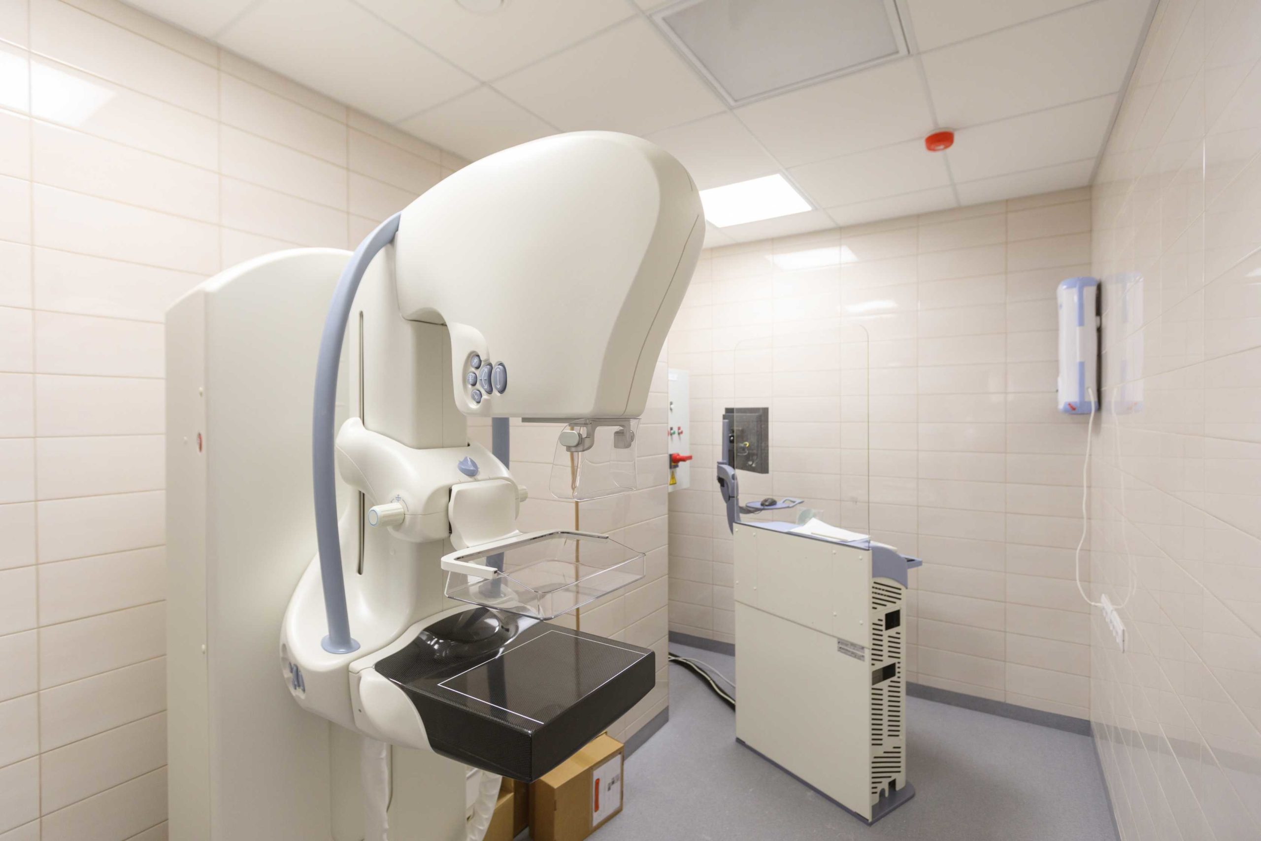
Thanks to technological innovations, the diagnosis and treatment of different forms of cancer have become more thorough, helping doctors improve patient outcomes. For example, 3D mammography provides more detailed imaging that shows tumorous growth from almost every angle—a vast improvement over the 2D imaging of the past.
Medical imaging’s advancement from 2D mammography to 3D mammography techniques was a real breakthrough in the area of breast cancer care. Although 2D images allow radiologists to detect abnormal breast tissue growth, they can also overlook abnormal tissue development that has replaced healthy breast tissue. As a result, breast tumors might go undetected and develop into malignant tumors.
The innovative 3D mammography technology allows radiologists to observe normal and abnormal breast tissue from various angles. The 3D mammography technique is used in conjunction with the digital (2D) mammography technique. However, 3D gives a more comprehensive view of a patient’s breast tissue.
Women who are at risk of breast cancer may exhibit any of the following symptoms:
If you experience any of these signs, see your doctor immediately to schedule a diagnostic mammography.
Early breast cancer detection provides better treatment options, improves the chances of survival, and enhances the quality of medical aid given to patients.
Diagnostic mammography is performed when patients feel any abnormal growth, pain, lump, or swelling in their breasts. Patients can discuss these abnormalities with their physicians and schedule a diagnostic mammography.
Compared to digital mammography, diagnostic mammography allows the radiologist to view the breast tissue from multiple angles. This enables physicians and radiologists to detect and diagnose any abnormal tissue growth.
Patients can discuss the results of their diagnostic mammography with our experienced breast radiologists.
How to prepare for a mammogram
Female patients can schedule a mammogram after it has been prescribed by their primary care physician or surgical specialist.
TOPS Comprehensive Breast Center is an outpatient facility that provides patients and physicians with best-in-class surgical care equipment. We’re committed to helping patients in our advanced facility, which is used by our trained and experienced doctors for surgical procedures to optimize patient outcomes.
We also accept major medical insurance plans for most procedures at our facility.
Get in touch with us to learn more about how we can help you.