3D mammography is a comprehensive radiological imaging technique that captures breast tissue images from multiple angles and is used in conjunction with 2D mammography.
The X-ray arm moves in an arc motion over and around the breast and captures images from different angles. These images are then fed into a computer and digitally compiled to create 3D, 1-millimeter-thick images of the breast tissue.
These 3D breast tissue images give a clearer picture and allow radiologists to detect, identify, and locate any abnormalities in the breasts.
Moreover, 3D mammography can help detect cancer in its early stages and lower the need for follow-up mammograms. Early detection, diagnosis, and treatment are the three most important factors in achieving favorable patient outcomes.
3D mammography is used in conjunction with the 2D mammography procedure. The procedures are performed simultaneously, so you don’t need to experience additional compression for this process.
Since the X-ray arm is capturing breast tissue images from multiple angles, the 3D mammography procedure can take slightly longer than a traditional 2D mammogram.
3D mammography is ideal for all women who are considering undergoing a general screening or diagnostic mammogram. Doctors and radiologists can get a clear image of the breast tissue. Moreover, 3D mammography helps detect breast cancer in its early stages.
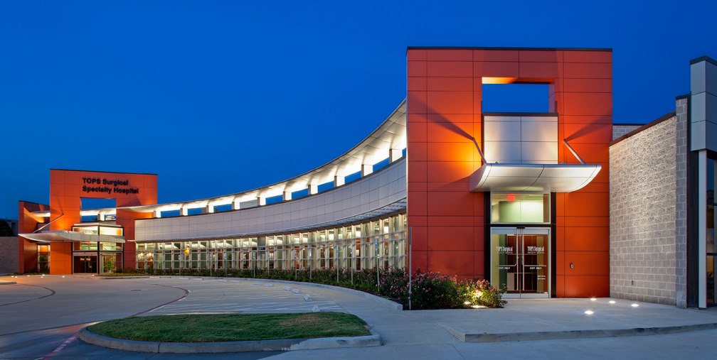
You can benefit from our 3D mammogram services at TOPS Comprehensive Breast Center. With the latest radiological mammography techniques and technologically advanced medical equipment, the center aims to streamline your medical treatment. Our healthcare staff and competent physicians are equipped with the knowledge and expertise needed to help you through your health journey.
You can get in touch with us to schedule your annual mammogram appointment at TOPS Comprehensive Breast Center. You can even request to see our list of doctors and learn how we can help you in the process.
Tomosynthesis is an advanced type of mammography imaging technique doctors use to screen for breast cancer in patients who may be in the early stages and showing no symptoms. Your doctor may use it as a diagnostic tool if your are showing symptoms of breast cancer symptoms.
Sometimes normal breast tissue can overlap and appear abnormal during a 2D mammography. This can prompt your radiologist to call you back for a follow-up session.
However, 1-millimeter-thick images offer a comprehensive and thorough view of the breast tissue. This helps reduce errors and allows radiologists to identify breast tissue abnormalities more confidently. A 3D mammography can capture the minutest details and help radiologists compile an accurate image of the breast tissue.
Screening mammograms are routinely performed to detect and identify any abnormalities in women who are not experiencing symptoms. A radiologist might recommend a callback for a diagnostic mammogram to rule out any suspicious abnormalities captured during a screening mammogram.
You will be exposed to a very low amount of radiation. The dose is almost the same as that of a traditional mammogram that is done on film. The radiation dose from a mammogram is much less than that from a CT scan or even a barium X-ray.
Your radiologist may recommend scheduling follow-up diagnostic mammograms to evaluate any abnormal breast tissue detected on your first mammogram. However, with our revolutionary 3D mammography, follow-up mammograms have been reduced up to 15%.

Phone: (281) 539-2900
Fax: (281) 715-4525
7 Day a Week 24 Hours a Day
Driving Directions